Dr. Szilvia Pellion, dermatologist, highlights the importance of self-exams, screenings, and prevention, sharing valuable tips that are great to keep in mind anytime.
What signs suggest it’s time to have a mole checked out?
If a mole changes—growing wider or taller, shifting color, itching, bleeding, hurting, developing irregular or uneven edges—you should show it to a dermatologist. Also, if a new spot appears on your skin, especially after a certain age, it’s wise to get it checked promptly.
What happens during an exam?
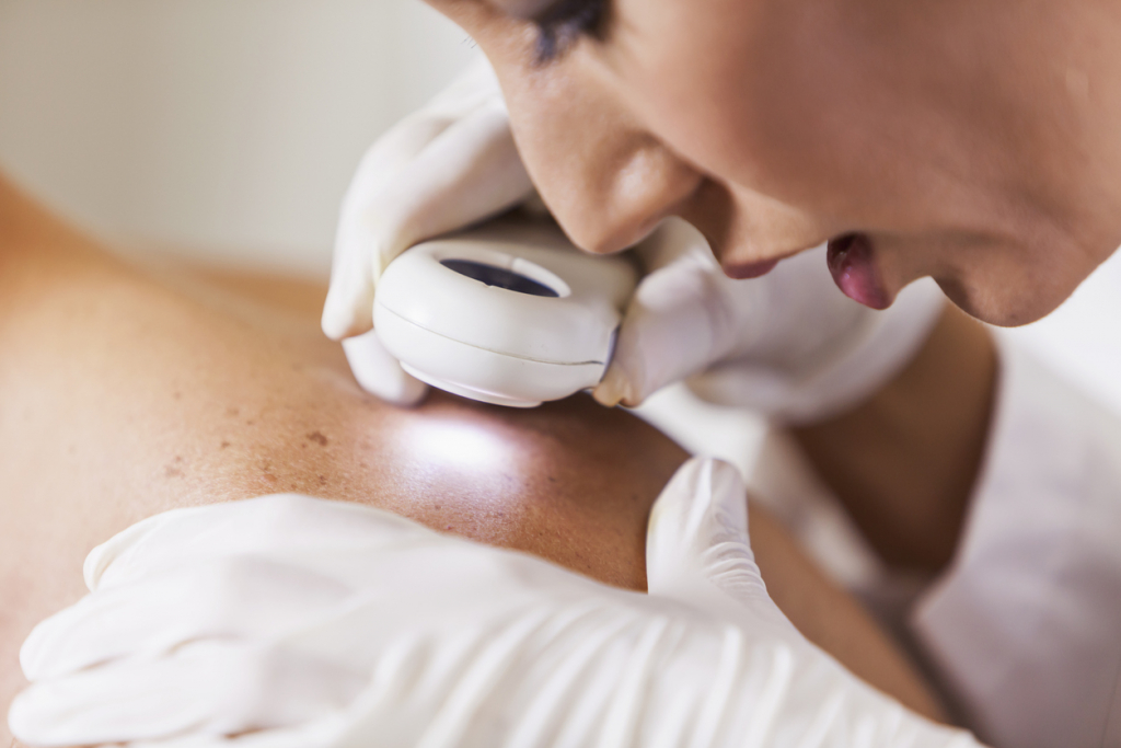
A handheld dermatoscope is used to examine your entire skin surface, checking each mole’s structure closely. Many clinics use special digital mole and skin cancer screening devices to capture and analyze images. For most patients, the handheld exam is enough, but if you have many irregular moles, a digital exam is highly recommended.
How often should you get a full skin check by a dermatologist?
Once a year is ideal. If you’ve had skin cancer, have many moles, or belong to a higher-risk group, checks every 3 to 6 months might be necessary.
What is melanoma, and when is it cancer?
Melanoma is a type of skin cancer that develops from pigment cells. It’s one of the most aggressive cancers, capable of spreading quickly, so early detection and removal are crucial. Around 20-30% develop from existing moles, but most appear as new growths on the skin. About 95% are brown, black, or gray and contain pigment, while 5% show up as red or light-colored spots.
What does stage one melanoma mean?
Stage one melanoma means malignant cells are only in the top skin layer (in situ melanoma) and haven’t spread deeper. At this stage, no metastasis is expected. If cancer cells reach the dermis or deeper layers, the Clark scale (five levels) and Breslow thickness help determine prognosis and if further treatment is needed.
Staging tells us how advanced the melanoma is. In stage one, cancer cells affect only the skin and aren’t found in nearby lymph nodes or other organs.
What follow-up care do melanoma patients need?
Patients diagnosed with melanoma are cared for in oncology centers, where their stage guides the type, frequency of exams, and treatments needed.
How can we tell the difference between a mole, an atypical mole, and melanoma?
Moles can be regular or atypical, showing various irregularities. Using the ABCDE rule and a dermatoscope, pigmented skin spots are scored to estimate whether a growth is benign or possibly malignant, like melanoma.
The final diagnosis always comes from histological and special immunohistochemical tests, which confirm the conclusive opinion.
The ABCDE rule:
- A for Asymmetry – how irregular is the mole’s shape, and has it changed?
- B for Border – are the edges sharp or blurred?
- C for Color – how many colors and shades does it have? Is it uniform or multicolored?
- D for Diameter – lesions larger than 5 mm (about 0.2 inches) have a higher chance of being melanoma.
- E for Evolving – has the mole changed over time in size, color, or surface?
What factors increase the risk of skin cancer?
High risk factors include lots of sun exposure, tanning beds, severe sunburns in childhood, family history of melanoma, previous skin cancer, skin types I and II, light hair and skin, freckles, many irregular moles, moles exposed to constant irritation or injury, changes in a mole, or large congenital moles.
How can we prevent skin cancer?
Get your moles checked yearly and perform regular self-exams at home. If you notice changes or new spots, see a dermatologist promptly—don’t delay. Avoid sun exposure during peak hours (11 a.m. to 3 p.m.), apply sunscreen daily, and never let your skin burn. Skip tanning beds—they’re even more harmful than sunbathing.
Should we remove any mole?
Moles are usually removed only when medically necessary—if they’re irregular, previously injured, constantly irritated, or if they bother the patient by protruding from the skin. Every removal is followed by a histological exam.
How is a mole removal done?
Mole removal is recommended with surgical excision under local anesthesia, always followed by histological analysis. The edges are stitched together, and stitches are removed between days 5 and 21, depending on the location of the mole. Learn more about mole removal here.